

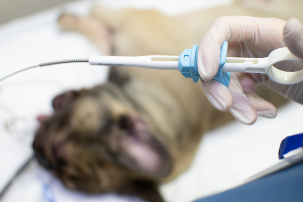
A new lump on your dog or cat can be a harmless fatty deposit or an early cancer, and neither sight nor touch can tell you which. The only reliable way to know is to sample the cells inside the mass. A fine needle aspirate uses a small needle to pull a few cells for the lab and usually delivers a quick answer with very little stress on your pet. A biopsy removes a piece of tissue and gives a fuller picture when the location or behavior of the mass calls for one.
At Oliver Animal Hospital in Austin, lump evaluation starts with a gentle nose-to-tail exam, in-clinic diagnostics, and a mobile specialist partnership for the cases that need more advanced care. Same-day urgent care appointments are available when a lump is changing quickly or when a worried family wants answers sooner. Our job is to explain what the findings actually mean and what realistic next steps look like for your pet. If you have found something on your dog or cat that you want a closer look at, book a visit with us.
The Short Version on Lump Testing
- No veterinarian can tell whether a lump is harmless or cancerous just by looking at it or feeling it, so sampling the cells is the only way to know for sure.
- Fine needle aspiration is the quick, low-stress first step for most masses, while a biopsy is reserved for masses that need a tissue-level look at grade, invasion, and margins.
- Some situations, like splenic masses and chronic GI disease in cats, skip aspiration entirely because they require tissue architecture to diagnose reliably.
- A benign result is good news, but it does not end the story, since new masses can appear and some known ones can change, which makes home monitoring worthwhile.
Why Can’t My Vet Tell What a Lump Is Just by Looking and Feeling?
No one can reliably diagnose a lump by sight or touch, not even a veterinarian with decades of experience, because malignant tumors and harmless growths can look and feel essentially identical. A soft, freely movable bump under the skin might be a benign fatty deposit or an aggressive soft tissue sarcoma, and the outside gives nothing away.
That is a hard thing to sit with when you have just found something new on your pet. Cancer in dogs and cats is common enough that roughly one in four dogs develops it at some point. Firmness, movement, size, and even how fast something grows can all point in the wrong direction. The needle, not the fingertip, is what settles it.
A lump is only one way trouble surfaces, and the broader signs of cancer in pets include unexplained weight loss, sores that refuse to heal, and quiet shifts in appetite or energy that are easy to write off. Any of those changes, with or without a visible lump, is worth a look. When something feels off, it’s better to sample early rather than watch and wait. Most of the time, the answer is reassuring, and getting it sooner spares you weeks of worry.
What Kinds of Lumps Do Dogs and Cats Actually Get?
Dogs and cats develop a whole cast of common lumps, and the gentlest and the most serious can feel exactly the same to the hand. A firm little bump could resolve on its own within weeks, or it could be one of the more aggressive tumors we see. Appearance alone never tells the whole story, and the only way to tell two masses apart is to look at the cells.
What Are the Most Common Lumps on Dogs?
Dogs grow a wide variety of skin masses that span the full range from completely harmless to genuinely concerning. Several different forms of skin cancer in dogs can surface as bumps that look nearly the same to the eye, which is why a raised spot cannot be sorted into safe or dangerous by appearance alone.
- Histiocytomas: These small, button-like growths show up most often in young dogs and frequently disappear on their own within a few months.
- Melanocytic tumors: These pigmented masses range from benign spots on haired skin to far more aggressive melanomas in the mouth or on the toes, and their color says nothing about how they behave.
- Cysts: Fluid- or material-filled cysts are usually harmless and can even drain on their own, though they can mimic a solid tumor until sampled.
- Mast cell tumors: Mast cell tumors are the great imitators, disguising themselves as a harmless fatty deposit or even an insect bite while quietly being one of the most common malignant skin cancers in dogs.
- Lipomas: Even the reassuring soft, movable lipomas cannot be confirmed by touch, since a fatty deposit and a sarcoma can present the same way.
What Lumps Do Cats Get?
Cats develop fewer skin masses than dogs, but a larger share of feline lumps turn out to be malignant, and they tend to hide until they are advanced. That combination is why any new lump on a cat deserves prompt attention rather than a wait-and-see approach.
- Basal cell carcinoma: Feline basal cell carcinoma often shows up as a firm, raised, sometimes pigmented nodule, and while many are locally contained, only sampling separates it from more dangerous look-alikes.
- Squamous cell carcinoma: Squamous cell carcinoma in cats often appears as a crusty sore on a white ear or nose that is easy to mistake for a scratch until it has already grown aggressive.
- Fibrosarcoma: A firm, deep-seated mass may be a fibrosarcoma in cats, an invasive tumor that tends to return locally if not fully removed, which makes tissue-level information especially valuable.
- Mast cell tumors: Feline mast cell tumors behave differently from the canine kind, often surfacing as small, firm nodules on the head or neck, and they can involve internal organs in ways no exam will reveal.
What Can a Fine Needle Aspirate Tell Me About My Pet’s Lump?
A fine needle aspiration is usually the first test we reach for because it tells us the biggest thing, whether a lump is benign or malignant, with the least stress on your pet. A thin needle collects cells, and answers come back within a few business days.
The procedure is quick and well-tolerated, and it usually needs no sedation. The needle draws cells from the mass, and those cells are sprayed onto a slide and stained. A pathologist then studies them to identify the cell type and whether it looks benign or cancerous, a process called cytology. For deeper masses near the belly or chest, ultrasound guidance steers the needle to the right spot, and our in-house laboratory and specialist-led ultrasound let us handle much of that workup without a long wait.
Cytology handles a lot. It reads fatty tumors, many mast cell tumors, certain round-cell cancers, and infections or inflammation quite well. What it cannot always do is show how cells are organized into tissue, and that gap is exactly what pushes some cases toward a biopsy. When loose cells are not enough to grade a tumor or plan surgery, tissue becomes the next step.
When Does My Pet’s Lump Need a Biopsy Instead of an Aspirate?
A biopsy becomes the answer when your pet’s mass needs a tissue-level view that loose cells cannot give, such as the exact tumor type, its grade, how deeply it invades, or whether surgery removed all of it. That level of detail is what guides treatment planning.
The real difference in cytology versus biopsy comes down to loose cells rather than intact tissue, and only that view shows how a tumor is arranged, how aggressively it grades, and whether it was fully removed. That examination, called histopathology, is what surgeons and oncologists rely on to plan care for the many types of cancer they see. Biopsies come in a few forms: a needle-core biopsy takes a thin cylinder of tissue, an incisional biopsy removes a small piece of a larger mass, and an excisional biopsy takes the whole thing at once. Because they all involve cutting into tissue, sedation or anesthesia is part of the plan.
Bypassing the Needle Entirely: Masses That Demand Tissue From the Start
Some masses bypass aspiration entirely because a reliable diagnosis depends on seeing how the tissue is built, not just what cells are present. In these situations, an aspirate can be misleading or even risky, so tissue is the starting point rather than a fallback. Two classic examples come up again and again: a mass on the spleen, and chronic digestive disease in an older cat.
Is a Splenic Mass Benign or Hemangiosarcoma?
A mass on the spleen is one of the hardest calls in the exam room, because imaging cannot reliably separate a benign growth from hemangiosarcoma in dogs, and the definitive answer often comes only after the organ is removed and the tissue examined. Aspirating a splenic mass is frequently unreliable because these growths sample poorly, and the spleen’s rich blood supply adds a real bleeding risk.
Because of that, we often recommend removing the spleen, a procedure called splenectomy, even before the diagnosis is fully confirmed. It feels backward to operate without the answer in hand, but for many of these masses, surgery is both the way to get the tissue and the treatment itself. The removed spleen goes to the lab, and histopathology delivers the definitive diagnosis that guides everything after.
Is My Cat’s Chronic Vomiting IBD or Lymphoma?
Distinguishing inflammatory bowel disease from small-cell intestinal lymphoma in a cat is one of the trickiest puzzles in feline medicine, because the two look nearly identical on every simpler test. In an older cat with chronic vomiting and weight loss, inflammatory bowel disease in cats and small-cell intestinal lymphoma look the same on signs, imaging, and bloodwork, so a full-thickness intestinal biopsy becomes the only way to point treatment in the right direction.
This matters because the treatment paths diverge sharply. One is managed with diet and anti-inflammatory support, while feline lymphoma of this type is a cancer that responds to a very different plan. Getting the biopsy is what keeps a cat from being treated for the wrong disease for months.
Reading the Report: The Three Outcomes and What Each One Means
Results generally land in one of three buckets, and each one points to a different next step. A benign result means we set up a monitoring plan, a malignant result opens a treatment conversation, and an inconclusive result guides which additional test comes next. Knowing which bucket you are in is what turns a scary unknown into a plan.
| Result | What it means | Typical next step |
| Benign | The mass is not cancer | Monitor at home, recheck as needed |
| Malignant | Cancer cells are present | Discuss surgery, staging, oncology, or comfort care |
| Inconclusive | The sample was not definitive | Repeat sampling or move to biopsy |
A malignant result is the one no family wants, but it is where having early, accurate information pays off most. Depending on the tumor, options can include surgery to remove it, staging tests to see whether it has spread, a referral for an oncology consultation, or a focus on keeping your pet comfortable. There is no single right answer, and we talk through what fits your pet and your family. When the kindest path is comfort-focused care, we offer compassionate support, and you can reach out to us to help you weigh a hard decision without pressure.
My Pet’s Lump Is Benign, So What Should I Watch for at Home?
A benign result is genuinely good news, but it does not close the file. New masses can show up alongside a known one, and some can change over time, so keeping an eye on things at home is how you catch those shifts early. It takes only a minute or two.
Once you know a lump is benign, note its size, shape, and location, and check it every few weeks. A few practical habits make this easy:
- Measure it: Use a ruler or a phone photo next to a coin so you can compare over time rather than relying on memory.
- Map it: Jot down where each lump sits, since a second or third one is easy to lose track of.
- Note changes: Watch for growth, a change in firmness, redness, discharge, or a lump that suddenly bothers your pet.
Call us if a known lump grows noticeably, changes texture, opens up, or seems painful, or if a brand-new mass shows up. Even a benign lipoma can eventually earn a recommendation for removal if it grows large enough to press on a joint or interfere with how your pet moves. If a lump changes suddenly, urgent care during our regular hours means you do not have to sit on it and wonder.
Catching Cancer in the Bloodstream Before You Feel a Thing
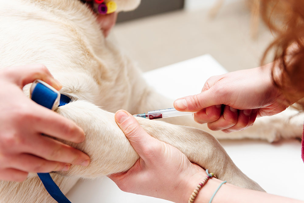
A newer category of blood test called liquid biopsy screening can detect genetic material shed by cancer cells into the bloodstream, sometimes before any lump is visible or felt. A positive result is not a diagnosis on its own, though. It is a signal that follow-up testing is needed to confirm what the blood picked up.
These cancer screening blood tests are most useful for pets at higher risk, particularly certain breeds. Some carry a heavier inherited risk than others, and the lymphoma risk in Golden Retrievers is a well-known example that makes proactive testing especially worth considering. This technology continues to evolve, and blood-based lymphoma screening is one avenue aimed at catching disease earlier than a physical exam ever could.
For senior pets and high-risk breeds, this kind of testing folds neatly into a regular checkup. We often talk it through during senior wellness visits, where an added blood draw can layer onto the workup already happening. It does not replace sampling a lump you can feel, but it can flag trouble that would otherwise stay hidden until later.
Frequently Asked Questions About Cytology and Biopsy for Pet Lumps
Does a fine needle aspirate hurt my pet?
Most pets tolerate an aspirate about as well as a routine vaccine. The needle is thin and the whole thing takes only a few seconds per sample, so sedation usually is not needed. Some pets feel a quick pinch and are more bothered by being held still than by the needle itself. For anxious pets or awkward locations, we can adjust our approach to keep the experience low-stress and calm.
Why can’t you just remove the lump and skip the testing?
Sometimes removing the whole lump is the right call, especially for a small, accessible mass. But taking one out without knowing what it is can backfire. If it turns out to be cancer, the first surgery may not have taken wide enough margins, which can mean a second, bigger operation. Sampling first tells us how much tissue needs to come out and whether staging should happen before anything is cut.
How long do results take to come back?
An aspirate sent for cytology usually comes back within a few business days, sometimes faster. Sometimes we can even do them in-house, depending on the lump type. A biopsy takes longer, often one to two weeks, because the tissue has to be specially processed before a pathologist can read it. We know the wait is the hard part, so we will let you know what to expect for your pet’s specific test and follow up as soon as the report is in.
Fewer Weeks of Wondering, Sooner
Testing a lump early works in your favor no matter how it turns out. The bump that comes back benign hands you relief and a simple monitoring plan, and the one that comes back malignant gives you far more treatment options when it is caught before it has had time to spread. Waiting rarely makes either outcome better.
If you have found something new on your pet, do not sit and wonder. Oliver Animal Hospital offers urgent care for dogs and cats in Austin when a lump is changing or a family just wants peace of mind. Whenever you are ready for a closer look, you can request a visit.






Leave A Comment