Do you have one of those adorable dog breeds with floppy, droopy skin? Maybe a flat-faced breed with cute squashed noses? Or perhaps just a pet who has recurring eye issues: squinting, redness, tearing, or chronic corneal ulcers? Certain breeds are predisposed to having eyelid issues, but they can occur in any pet. Knowing what to watch for, and if your pet is at risk, is the first step.
Entropion and ectropion are structural eyelid conditions where the lid itself is the problem, either rolling inward against the eye surface or drooping outward and leaving the inner tissue exposed. Both are painful, both worsen over time, and the good news is that both are correctable with surgery.
At Oliver Animal Hospital, we approach every case with gentle, compassionate handling to minimize stress, especially for pets with painful eyes. Our team can evaluate your pet’s eyelid anatomy, determine whether the condition requires medical management or surgical correction, and walk you through every step of the process. Request an appointment or contact us if your pet’s eye symptoms are not resolving with standard treatment.
Entropion vs. Ectropion: What Each Condition Actually Does
These two conditions sound similar but work in opposite directions, and the distinction matters for how they are managed.
Entropion occurs when the eyelid rolls inward, causing the fur and lashes along the lid margin to rub against the cornea (the clear outer surface of the eye) with every single blink. The friction is constant and causes significant pain, chronic tearing, redness, and over time, corneal scarring and ulceration that can impair vision. Entropion can be present from birth as a hereditary condition or can develop gradually as a pet ages.
Ectropion works the opposite way: the lower eyelid droops or sags outward, exposing the inner eyelid (conjunctival) tissue to air, dust, pollen, and bacteria. Rather than causing friction, ectropion causes chronic low-grade inflammation and infection because the eye’s natural protective mechanisms no longer work properly. The exposed tissue collects debris and stays dry instead of being kept clean by normal blinking.
Some pets develop both conditions in different parts of the same eyelid, a combination called diamond eye, which requires attention to both components. The broader range of eye conditions affecting pets includes many overlapping signs, which is why accurate diagnosis is the essential first step.
Which Pets Are Most at Risk?
Genetics and facial structure drive most eyelid abnormalities. Certain breeds are significantly overrepresented, and knowing your dog or cat’s predispositions helps with early detection.
Breeds commonly affected by entropion:
- Chow Chows, Shar-Peis, and Mastiffs (heavy skin folds)
- Golden Retrievers, Labrador Retrievers, and Rottweilers
- Bulldogs, Pugs, and French Bulldogs, where flat facial structure causes medial canthal entropion at the inner corner of the eye
- Basset Hounds, St. Bernards, and Great Danes are prone to ectropion
Eyelid disorders across breeds vary in severity and onset. Some puppies are born with visible entropion; others develop it gradually as they grow. Hereditary eyelid conditions often become apparent in the first year of life.
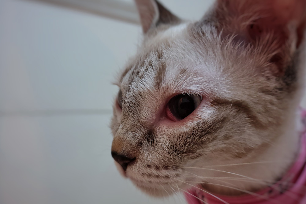
In cats, flat-faced breeds including Persians and Himalayans are prone to entropion due to their compressed facial structure. The inner corner of the eye is particularly affected in these breeds, and the condition often develops alongside other ocular surface changes.
Environmental factors and chronic eye disease can also cause acquired entropion in any breed, particularly in older cats where long-standing eye irritation causes the lid to roll inward over time.
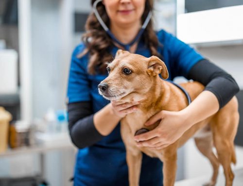
Regular veterinary wellness care is where breed-specific eye concerns get flagged early, before they cause significant damage.
Signs That Your Pet’s Eyes Need Attention
Eyelid conditions cause real pain, and the signs reflect that, even when pets try to mask discomfort in their typical fashion.
Warning signs that warrant an eye exam:
- Excessive tearing or watery discharge that keeps returning
- Squinting, keeping one eye partly closed, or favoring one eye in bright light
- Pawing, rubbing, or scratching at the face or eye
- Red or pink inner eyelid tissue visible at the corners
- Cloudiness, haziness, or color change in the cornea
- Thick or discolored discharge, or crusting that recurs after cleaning
- A visibly drooping lower lid or a lid that appears rolled in
Signs of eye pain in pets are not always obvious. A pet that stops wanting their face touched, avoids sunlight, or seems irritable when approached near the head may be experiencing significant eye discomfort. Do not dismiss these behavioral changes as unrelated.
If you notice any combination of these signs, an eye examination is the right next step. Contact us to schedule an evaluation during our open hours (Monday through Friday at Oliver Animal Hospital in Austin).
What Happens Without Treatment
Untreated entropion creates escalating damage. Lash friction on the cornea causes corneal ulcers, which are painful open wounds on the eye surface that can become infected, deepen, and in serious cases perforate. Scar tissue from chronic irritation permanently clouds the corneal surface, reducing vision even after the structural problem is corrected.
Untreated ectropion leads to recurrent conjunctivitis, chronic eye discharge, drying of the sensitive eye structures, and increasing susceptibility to infection as the exposed tissue loses its normal protective barrier.
Neither condition improves on its own. The earlier the structural problem is identified and addressed, the less secondary damage accumulates.
How We Diagnose Eyelid Conditions
A thorough ocular examination evaluates multiple components:
- Visual inspection of lid position: assessing where the lid margin sits relative to the eye and whether it contacts the corneal surface
- Numbing drops: applying topical anesthetic before examination allows full evaluation of lid position without pain-related squinting interfering
- Tear production measurement (Schirmer tear test): assesses whether the eye is producing adequate tears, since dry eye often coexists with or mimics entropion
- Fluorescein staining: a harmless orange dye applied to the eye reveals corneal ulcers or surface damage invisible to the naked eye
- Magnified evaluation: allows close inspection of the lid margin for eyelash problems including distichiasis (lashes growing from abnormal follicles) and trichiasis (normal lashes redirected toward the cornea), which can mimic entropion
- Facial conformation assessment: evaluating the overall facial structure helps distinguish true entropion from lash-related friction
The full picture from these tests guides whether the condition requires surgery, medical management, or a combination approach. Our services include the diagnostic tools needed to evaluate your pet’s eyes thoroughly in a single visit.
Treatment Options: From Temporary Tacking to Permanent Repair
Temporary Eyelid Tacking
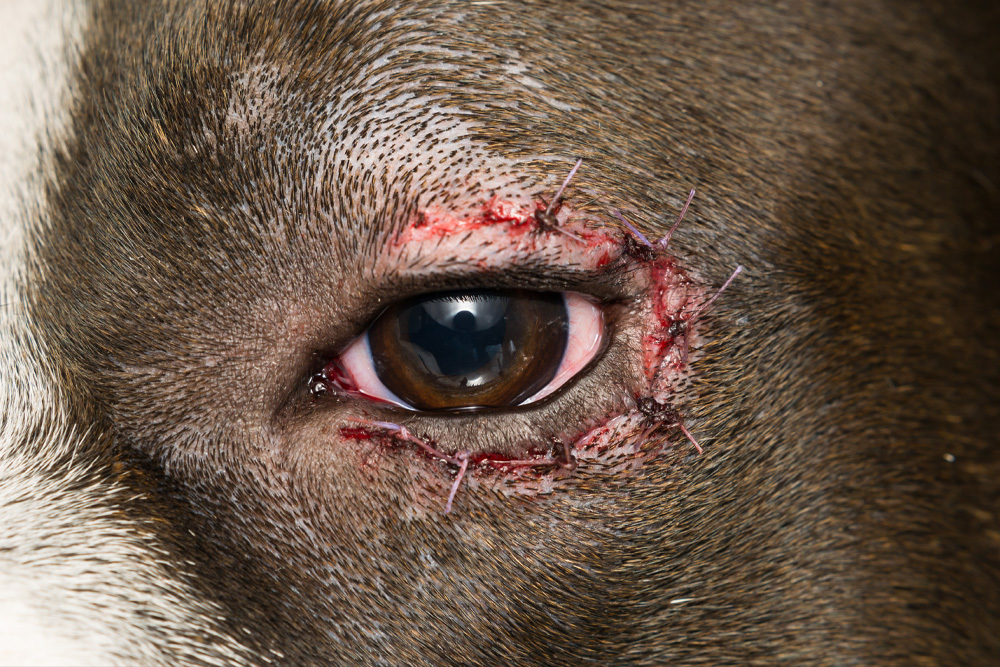
In young puppies where entropion is present but facial growth is not yet complete, temporary eyelid tacking uses small, strategically placed sutures to hold the eyelid in a corrected position. The goal is to protect the cornea during the period when the final eyelid position is not yet determined. Some puppies resolve after tacking as their facial structure matures; others require permanent surgery once growth is complete. Tacking gives young pets a chance to develop normally without accumulating corneal damage in the meantime.
Permanent Surgical Correction
Definitive eyelid surgery is performed when the condition is clearly structural and the pet has finished growing. For entropion, the procedure involves removing a precise strip of skin and tissue to roll the lid margin back into its correct position. For ectropion, the technique tightens and repositions the drooping lid. The exact approach varies based on the pet’s specific anatomy, the location and degree of the problem, and species.
Precision matters enormously in eyelid surgery. Overcorrection of entropion can create ectropion, and undercorrection leaves the original problem unresolved. This is why careful surgical planning and technique are essential to good outcomes.
Entropion in Cats
Entropion in cats often presents differently than in dogs. Rather than appearing at birth as a hereditary condition, feline entropion frequently develops later in life, often secondary to chronic eye surface disease or as a consequence of long-standing squinting. Treating the underlying eye disease is often necessary alongside correcting the lid position. Cats also tend to require smaller, more delicate corrections than dogs.
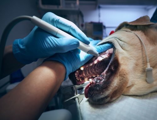
What Surgery Day Looks Like
Walking into a surgical procedure with a clear picture of what to expect reduces anxiety for both you and your pet.
The day of surgery:
- Pre-anesthetic assessment: bloodwork may be recommended, particularly for older pets, to confirm safe anesthetic risk
- Customized anesthesia protocol: tailored to your pet’s age, size, and health status
- Pain management before the procedure begins: pre-operative medications ensure your pet wakes up comfortable, not catching up to pain
- Continuous monitoring throughout: heart rate, breathing, blood pressure, and temperature are tracked by dedicated staff
- Precise surgical technique: careful marking, measurement, and suture placement are what produce a symmetric, well-positioned result
Our team at Oliver Animal Hospital treats every pet in our care like they belong to us. That means the same careful attention to anesthesia and pain management that we would want for our own animals.
Recovery: What to Expect
The First Several Days
Normal and expected:
- Mild swelling and bruising around the eye, peaking around 24 to 48 hours
- Small amount of clear discharge
- Sleepiness and reduced energy from anesthesia and pain medication
Signs to call about:
- Rapid or significantly worsening swelling
- Thick yellow or green discharge
- Bleeding at the incision site
- Sutures that appear loose or missing
Keeping the e-collar on at all times during recovery is not negotiable. The instinct to paw at a healing eye is strong, and removing the cone, even briefly, is often when sutures are lost or wounds are reopened.
Administering eye medications at home can feel intimidating at first. Our team will walk you through the technique before your pet goes home, and we are available to answer questions during recovery. Contact us with any concerns rather than waiting for the recheck.
Keeping the eyelid area clean between medication doses is also important. Optixcare eye wipes are a gentle option for removing discharge and debris around the healing area without disturbing the sutures.
The Healing Timeline
- 10 to 14 days: sutures are typically removed at the recheck appointment; initial healing is largely complete
- Weeks 2 to 6: final eyelid position settles as residual swelling resolves; this is when the full correction becomes visible
- Follow-up exams: confirm proper healing and allow early identification of any areas that may benefit from minor revision
Most pets show obvious relief within the first day or two after surgery. The squinting, pawing, and chronic tearing that drove the evaluation typically improve dramatically once the lid is in its corrected position.
What Outcomes Look Like
Success in eyelid surgery means a lid that sits in a normal position without causing friction or leaving tissue exposed, and an eye that is comfortable, clear, and no longer accumulating damage. Most pets achieve this with a single well-executed procedure.
Factors that influence outcomes:
- How much secondary damage (corneal scarring, ulceration) was present before surgery
- Whether the eyelid correction achieves proper position without over- or undercorrection
- The pet’s age and how long the condition was present before treatment
- Cone and medication compliance during recovery
Our team performs these procedures with the same level of care we would expect for our own pets, and we follow each case through recovery to make sure the result is what it should be.
Frequently Asked Questions
Is eyelid surgery necessary, or can eye drops manage the condition?
Drops can temporarily relieve symptoms and are sometimes used short-term to protect the cornea while planning surgery. They do not correct the underlying structural problem. If the lid physically contacts the cornea, the friction continues regardless of what medications are used.
Can entropion or ectropion come back after surgery?
In most cases, surgical correction is permanent. Occasionally, a pet requires minor revision if healing does not produce perfect symmetry or if the condition was severe enough to need a staged correction. This is discussed at the pre-surgical consultation based on the individual case.
At what age can a puppy have permanent eyelid surgery?
Generally, permanent surgery is delayed until growth is complete, typically six months to a year of age depending on breed. Temporary tacking is used in the meantime to protect the cornea.
Are both eyes usually affected?
Many cases are bilateral (affecting both eyes) because the underlying cause, whether genetics or facial structure, affects both sides. Each eye is evaluated and, if needed, corrected.
Helping Your Pet See Comfortably Again
Entropion and ectropion are painful, progressive, and entirely correctable when addressed with proper evaluation and surgical technique. Pets that live with these conditions untreated are uncomfortable every day. Those that receive timely correction typically experience fast, dramatic relief and go on to have normal, comfortable eye function.
If your pet has been squinting, tearing, or showing other signs of eye discomfort, request an appointment at Oliver Animal Hospital in Austin or reach us at (512) 892-1000. The sooner the eyelid anatomy is evaluated, the sooner your pet gets relief.
Leave A Comment